Release date: 2017-07-27

The liver is a particularly "slippery" organ that adds a lot of difficulty to liver tumor surgery. Because the doctor found a life-threatening tumor through a CT scan, the patient's position may have "slid" an inch when the patient was lying on the operating table. The liver is an important organ of blood circulation in the human body. The vicinity of the tumor may be an important blood vessel. This fluidity is undoubtedly a challenge for successful surgery.
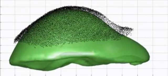
The doctor can gently depict the exposed liver surface with a special stylus, capturing the shape of the liver during surgery, and the computer can match the CT scan image. This GPS-like ability is far better than guessing where the tumor and blood vessels are, but even this roadmap may have centimeter-level deviations, and the rest can only rely on the doctor's speculation. 
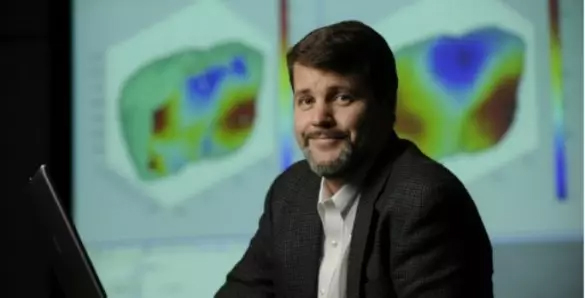
Michael Miga Image credit: Vanderbilt University
Michael Miga, a professor of biomedical engineering at Vanderbilt University, and his team offer a promising solution: with tracking tools, surgery-testing software can better match CT scan images. This is an advancement that can help more than 500,000 liver patients worldwide each year. Their article "Deformation Correction for Image Guided Liver Surgery: An Intraoperative Fidelity Assessment" was published this month in the magazine "Surgery."
The Abdominal Surgery Pathfinder Stylus System was sold to Analogic Corporation in 2014 and is used at top cancer centers. Miga, the developer of the system, pointed out that "the shape of the liver in the diagnostic imaging of the body and the shape of the surgery are very different," he said. "If you try to find a dime-sized tumor and avoid the blood vessels. Then you need to avoid the error. But the difficulty is that the doctors have so far been able to evaluate the operation of an organ. The GPS spectrum of CT navigation is only in centimeters. This is very dangerous, especially the cutting position is mainly from the main position. The blood vessels are very close."
If you want to find a way to fix this error without further investment in expensive equipment, you can use computer software to make the computer model start from the original shape of the liver and simulate the external stress in the surgery—for example, wrapping the liver in Lift up in the gauze. The computer can adjust the GPS spectrum of the CT navigation to better match the shape of the organs exposed during the procedure.
A randomized, double-blind study of 20 subjects enrolled at the Memorial Sloan Kettering Cancer Center in New York. In the study, the doctor will display 6-7 CT images for a total of 20 patients with liver tumors in the operating room, for a total of 125 images. The CT map will be aligned with the original Pathfinder or Miga's new enhanced CT image to correct for distortion. The doctor does not know which one is being displayed. They use the touch to touch the patient's liver and observe the display to evaluate the image calibration. The doctor then gives a correlation score of +3 to -3 for the image in the display. The display order is random, so it can be from original to enhanced, from enhanced to original, or left unchanged. The surgeon was able to correctly detect 73% of the changes in 125 different assessments.
Current surgery forces doctors to use a combination of science and labor to find a tumor and decide how much liver to remove to self-recover. This new deformation correction technique can be integrated into an image guided surgical system. Miga said that their new research has made a historic contribution to soft tissue abdominal image-guided surgery.
Reference material
[1] New tools help surgeons find livertumors, not nick blood vessels
[2] Software Models Liver's Movement toGuide Surgeons to Target Tumors
Source: Health New Vision
Preservation tubes are swabs with disposable virus sampling tubes to collect DNA tests for disposable nasal flocking sterile medical transport.
Made of high-quality PP material, high chemical stability, high temperature sterilization, suitable for multi-channel pipettes and automatic machines. Square well, U-shaped bottom design ensures minimal sample residue. Molded letters and numbers for easy positioning. Complies with SBS regulations and can be stacked stably. Good sealing performance, the top of the plate is flat and uniform, maintaining an effective seal. Optional gamma radiation sterile, DNase and RNA enzyme free, pyrogen-free.
Stable performance, low price, the material has a strong electrostatic adsorption capacity, can provide customized plastic card color according to user requirements, good color consistency
Non-invasive, painless and easy to operate. Samples are non-contagious and can be transported safely.
Disposable sampling swabs,Sterile sampling swab,Medical grade disposable swab
Jiangsu iiLO Biotechnology Co., Ltd. , https://www.iilogene.com