Rapid, efficient, and reliable early prediction of toxicity in vitro is critical for drug development and reducing the risk of drug clinical trials. Utilizing the new progress of modern life sciences, the establishment, application of new technologies, new methods and new models for preclinical safety evaluation and toxicological mechanism research of new drugs has become a new trend in the development of new international drugs.
The High Connotation Screening (HCS) system is an automated, high-resolution microscope that performs high-throughput analysis of changes in cell morphology or biochemical properties. With the help of automated HCS, pharmaceutical companies can quickly screen libraries of toxic compounds, which is a necessary step in the development of new drugs. As technology continues to evolve, HCS systems are becoming faster, more flexible, and more sensitive.
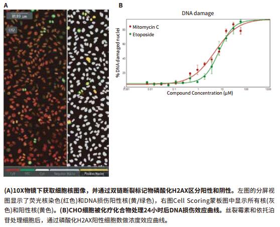
The latest DNA damage detection method - application of ImageXpress Nano imaging analysis system
DNA or chromosome damage is often involved in research because DNA damage is closely related to the occurrence of many diseases, such as genetic diseases and tumors. Radiation radiation, environmental effects, or combined drugs can cause DNA damage. Previous studies have shown that the phosphorylation site of the labeled histone H2AX on serine 139 is a sensitive method for early prediction of DNA double-strand breaks. In this paper, the experimental method of detecting DNA damage by automatic cell imaging is described in detail. The cell sample is treated by small molecule compound and imaged by phosphorylated histone H2AX immunofluorescence staining. In addition, three cell lines, CHO, Hela and PC-12, were added to the experiment, and mitomycin and etoposide were added to detect the effects of the compounds.
Advantage
- After the cells are fixed, the nuclei are fluorescently stained by immunoassay and imaged.
- Accurate calculation of DNA double-strand break sites
- On-the-fly analysis yields calculation results in real time
- Display positive holes by heat map
View full story

Live cell evaluation of cell cycle inhibitors
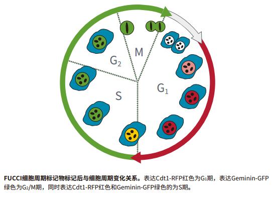
Monitoring the effects on cell cycle changes plays an important role in tumor development and drug discovery. For example, compounds known to inhibit mitosis are mostly used to slow the growth of tumor cells. Screening for high content of living cells has been validated to distinguish each cycle of cells. This technology, in combination with the BacMam transfection system, allows cells to express two cell cycle-associated fluorescent fusion proteins.
The delayed monitoring experiment lasted 2-3 days and the cells were placed in the environmental control chamber of the ImageXpress ® Micro high-content imaging and analysis system. The entire experiment maintained the normal growth environment of living cells. Live cells and fluorescent live cell images are acquired simultaneously at pre-set intervals. This complete protocol consists of a pre-built module that analyzes time-lapse images, identifies all cells based on brightfield images, and differentiates between cell cycles based on fluorescent protein expression.
Advantage
- Live cells continue to grow in the instrument for up to 72 hours
- Reduce experimental operation time
- Identify cells without labeling in bright field
- Detection of cyclical changes using cell cycle transfection reagents
View full story

High-intensity 3D toxicity analysis using hepatocyte spheres derived from pluripotent induced stem cells (iPSC)
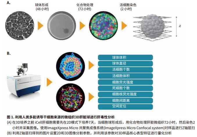
In developmental biology and tissue biology, 3D cell ball modeling can accelerate the process of transformational research. In vitro culture, if the cultured cells are added to the micropores of the round bottom, these enriched cells form discrete spheres. These discrete spheres contain both surface-exposed and deeply buried cells, proliferating and non-proliferating cells, an outer oxygen-rich layer and an internal anoxic center. Based on the above characteristics, compared with the traditional two-dimensional cell culture method, the sphere can better simulate the behavioral characteristics of tissues and individuals. Therefore, it is getting more and more people's attention. How to perform higher-throughput quantitative analysis of 3D samples has become a hot research topic.
In this example, MD has established and optimized an analytical method for confocal imaging and toxicity assessment of human pluripotent stem cell-derived 3D hepatocyte spheres.
Advantage
- Using human pluripotent to induce stem cell-derived hepatocytes to form hepatocyte spheres
- Evaluation of hepatotoxicity in 3D models screened in vitro
- Identify and segment target samples during 3D image analysis to achieve optimal segmentation

High-throughput screening for neurotoxicity
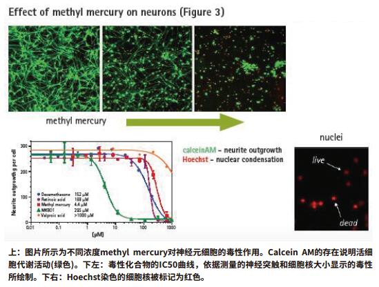
The nervous system is very sensitive to many toxic compounds and harmful substances produced in the natural environment. During the course of the disease, neurotoxicity can cause transient or persistent damage to the brain or the peripheral nervous system, such as spinal cord injury, stroke, traumatic brain injury, and the like. At the same time, this neurotoxicity is also the main cause of many neurodegenerative diseases such as Alzheimer and Parkinson.
High-throughput imaging techniques that induce human pluripotent stem cells can be used to screen for neurotrophic, protective or neurotoxicity of drug candidates or environmental pollutants. This section explains how to combine iPSCs, molecular instruments and software to automatically screen endpoints for pluripotent stem cells and neurotoxicity of live cells.
Advantage
- Fully automated image acquisition and analysis
- Quickly get multiple phenotypic parameters for each cell
- Real-time monitoring of the toxicity of living cells, which can last from a few minutes to a few days


Up-right Researching Biological Microscope
Up-Right Researching Biological Microscope,Discovery Microscope Lab,Labline Microscope,Medical Laboratory Microscope
Ningbo ProWay Optics & Electronics Co., Ltd. , https://www.proway-microtech.com